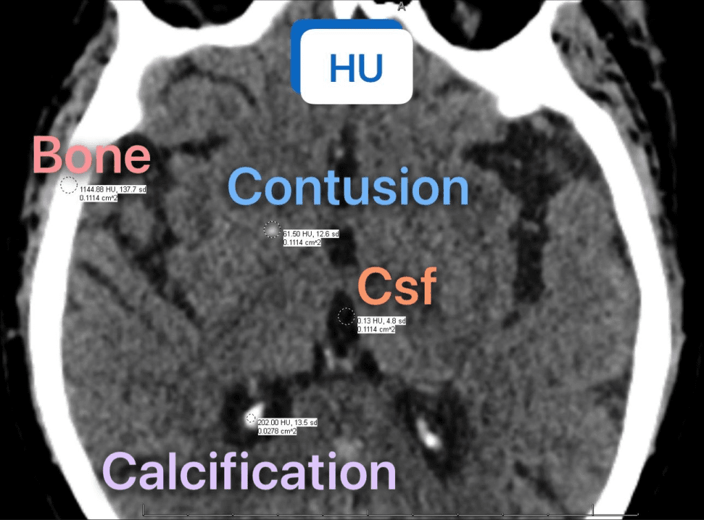
In emergency medicine, rapid and accurate interpretation of CT scans is crucial. Hounsfield Units (HU) provide a standardized scale to differentiate between various brain tissues and pathologies:
• Cerebrospinal Fluid (CSF): ~15 HU
• White Matter: +20 to +30 HU
• Gray Matter: +37 to +45 HU
• Acute Blood (Hemorrhage): +50 to +75 HU
• Cortical Bone: +700 to +1900 HU
• Calcifications: Typically >100 HU
Recognizing these values aids in distinguishing normal anatomy from pathological findings, such as differentiating calcifications from hemorrhages. For instance, calcifications often present with HU values exceeding 100, assisting in accurate diagnosis.