“Inferior… But Not Alone: When STEMI Spreads to the Back and Right Heart”

“Inferior… But Not Alone: When STEMI Spreads to the Back and Right Heart”

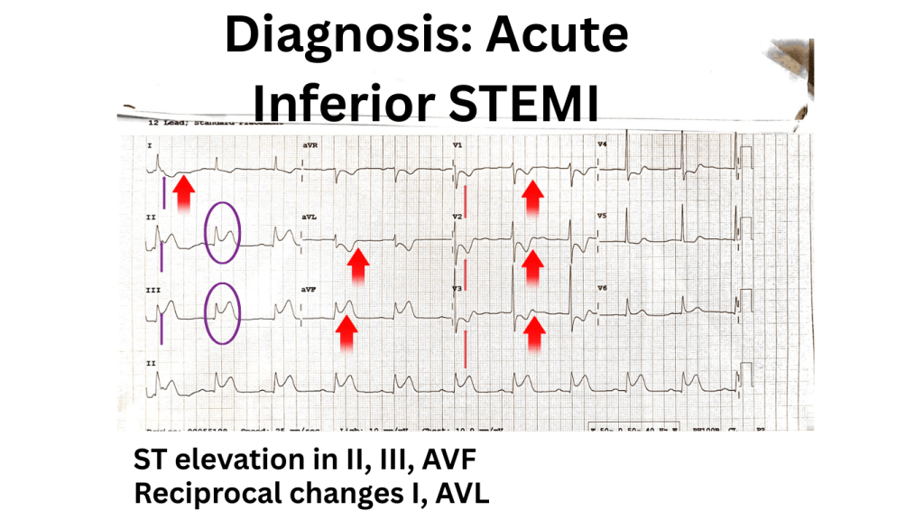
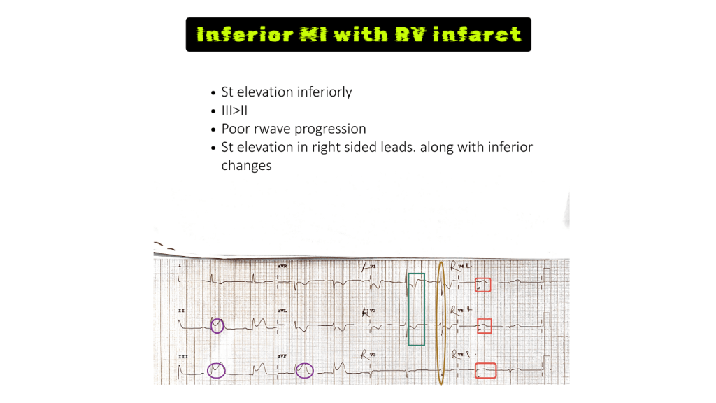
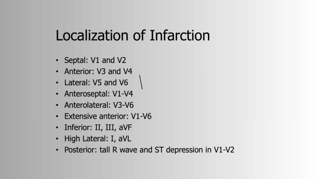
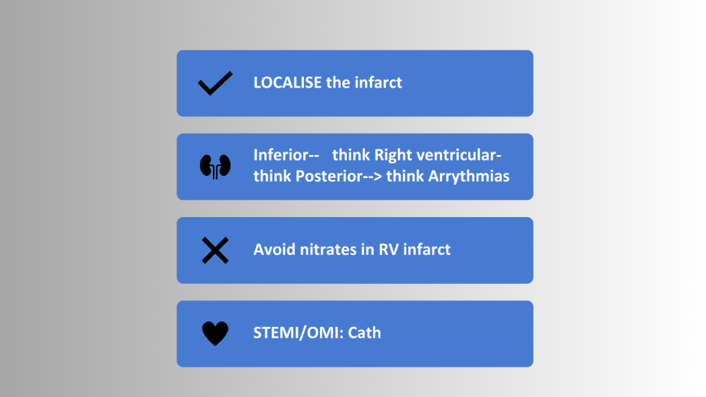
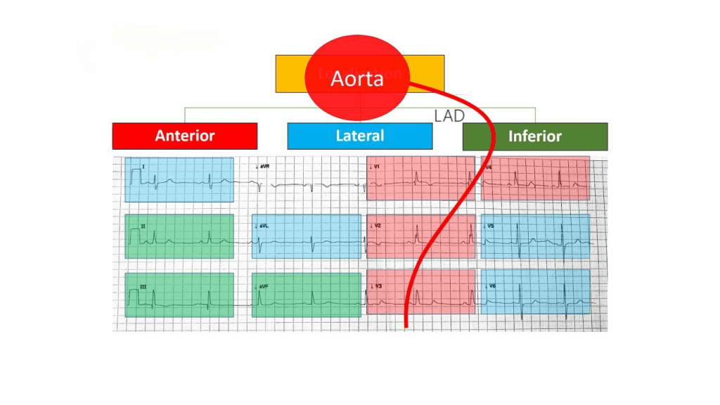
This case presents a 51-year-old male with an acute inferior ST-Elevation Myocardial Infarction (STEMI), diagnosed by ST elevation in leads II, III, and aVF. The key takeaway is that with any inferior STEMI, it is crucial to investigate for both Right Ventricular (RV) and Posterior wall involvement.

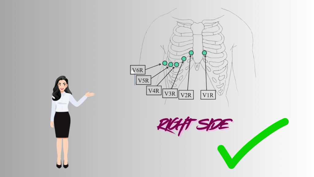
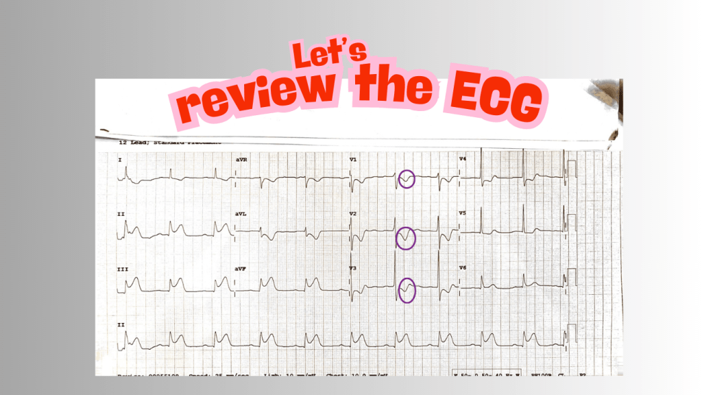
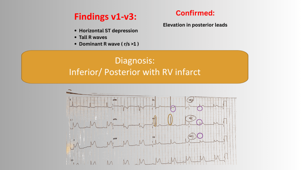
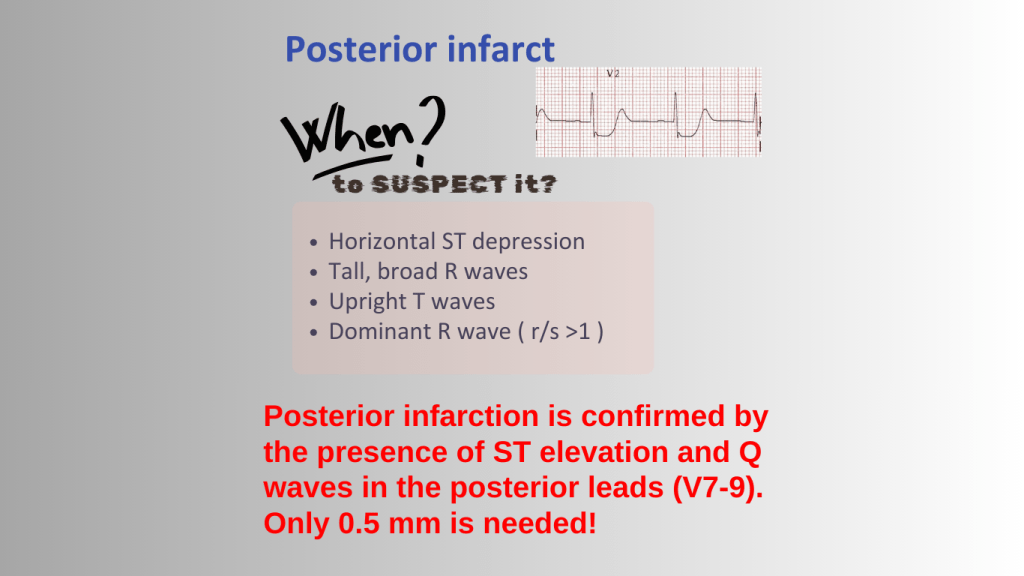
RV infarction is suspected when ST elevation is greater in lead III than lead II and is confirmed with a right-sided ECG. Posterior infarction is suspected with ST depression and tall R waves in leads V1-V3 and confirmed with a posterior ECG. Identifying an RV infarct is critical because nitrates must be avoided to prevent severe hypotension.

Read the story :

Title page of an ECG capsule prepared by Dr. Sabrina Berdouk, peer-reviewed by Abdolghader, featuring the EM Mastery Academy logo.
Illustration of a 51-year-old Sri Lankan male experiencing chest pain rated 5/10, with symptoms of nausea and no past medical history.
Illustration of an ECG machine displaying heart rhythm, with a printed ECG graph beside it.
12-lead ECG showing ST elevation in leads II, III, and aVF, suggesting an inferior STEMI.
Electrocardiogram (ECG) showing diagnosis of acute inferior STEMI with ST elevation in leads II, III, and aVF, highlighted by arrows, along with reciprocal changes in leads I and AVL.
Text explaining inferior infarct and its implications for patients with STEMI.
Illustration depicting the concept of Right Ventricular Infarct, featuring a character in a suit listening intently with the text 'What type of ECG do you ask next?'
An illustration showing the proper placement of ECG leads on the right side of a human chest with a woman pointing to the diagram.
Electrocardiogram (ECG) showing inferior ST elevation and indications of right ventricular (RV) infarct, including lead comparisons and annotations.
Infographic on RV infarction, highlighting signs to suspect it: inferior STEMI, ST elevation in v1, and greater ST elevation in lead III than II on 12 lead ECG. Includes note to confirm with right-sided ECG.
Image illustrating the importance of Right Ventricular (RV) involvement in myocardial infarctions, featuring the words 'Nitrates' with a caution sign and 'Arrhythmias' alongside a graphic of a heart.
Illustration depicting a medical scenario with healthcare professionals examining a patient. The text overlay reads, 'WHAT?! type of ECG do you ask next?'
ECG printout showing leads I, II, III, aVR, aVL, aVF, V1 to V6 with annotations highlighting areas of interest.
Diagram illustrating ECG leads for detecting posterior myocardial infarction, labeled with V4, V5, V6, V7, V8, and V9.
ECG findings for inferior and posterior myocardial infarction showing horizontal ST depression and tall R waves in leads V1-V3, confirming elevation in posterior leads.
Educational slide on posterior myocardial infarction diagnosis, detailing signs such as horizontal ST depression, tall broad R waves, upright T waves, and the dominant R wave. Includes an ECG image with V2 lead presentation.
Slide detailing the localization of myocardial infarction indicating specific ECG leads for different types of infarction.
Graphic summarizing key considerations for diagnosing and managing inferior ST-Elevation Myocardial Infarction (STEMI), including localization of the infarct, potential right ventricular involvement, and warnings against using nitrates.
Diagram illustrating the relationship between the aorta and different myocardial regions, including anterior, lateral, and inferior sections, with associated ECG readings.

Discover more from EM Mastery Academy

Subscribe now to keep reading and get access to the full archive.

Continue reading

Discover more from EM Mastery Academy

Subscribe now to keep reading and get access to the full archive.

Continue reading