
Think “worst chest pain of life” with neuro signs, weird vitals, or something just not adding up. That’s your trigger. If you’re not thinking about dissection when you should, you won’t diagnose it in time.
🚨 CASE 1: The Missed MI
A 62-year-old male with HTN and a 20 pack-year smoking history presents with “ripping chest pain.” It started suddenly, peaked immediately, and radiates to his back.
Vitals:
- BP 182/90 (right arm), 138/80 (left arm)
- HR 101
- ECG: Inferior ST elevations
- Troponin: Slightly elevated
What you think: STEMI.
What it is: Type A dissection compressing RCA → inferior STEMI pattern.
🎯 Pearl: If ST-elevation + back pain + BP discrepancy = assume dissection until proven otherwise. Do not give heparin until CT aorta rules it out.
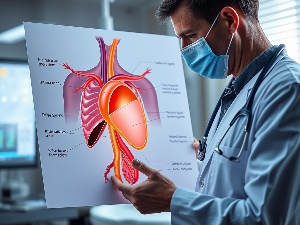
THE BASICS: What Actually Happens in Aortic Dissection?
- The intima tears, blood punches through into the media, and forms a false lumen.
- This false channel may cut off branch vessels, rupture, or compress the true lumen → ischemia, tamponade, death.
🧬 Variants you need to remember:
- Classic dissection: with flap and true/false lumen.
- Intramural hematoma: blood within wall, no flap.
- Penetrating ulcer: erosion of plaque through wall.
🎓 Treat them all as aortic catastrophes unless proven otherwise.
🔍 CASE 2: The Silent Neurologic Presentation
A 48-year-old woman presents with sudden-onset left-sided weakness. No chest pain. BP 210/95. No pulse in left radial artery.
CT head: Normal.
You’re about to call a stroke alert, but… something’s off.
CTA head/neck shows: no ICA occlusion.
CTA chest reveals: Type A dissection occluding left subclavian + left carotid.
🎯 Pearl: Not all dissections hurt. Some stroke patients have painless aortic dissection. Always check bilateral BPs, pulses, and neuro exam.
🔬 QUICK DIAGNOSTIC TOOLKIT
| Tool | Usefulness | Pearl |
|---|---|---|
| CT Angio Chest/Abd/Pelvis | 🥇Gold standard | Get it urgently if suspicion high |
| POCUS | Fast triage | Pericardial effusion? Aortic root dilation? |
| CXR | Non-specific | Widened mediastinum in ~60% |
| D-dimer | Limited | Cannot rule out dissection (especially intramural hematomas) |
| ECG | Can mimic MI | Normal ≠ normal. Watch for ischemia from RCA involvement. |
| Troponin | May be ↑ | Seen in up to 18% of AD cases |
🩺 HOW DO THEY PRESENT? RED FLAGS TO REMEMBER
- Pain
- Sudden, maximal at onset
- “Ripping,” “tearing,” or just “weird”
- Migrates down chest, back, abdomen, groin
- BP/pulse difference
- ≥ 20 mmHg between arms
- Absent or weak pulse in any limb
- Neurologic signs
- Stroke
- Syncope
- Hoarseness (recurrent laryngeal involvement)
- Spinal cord ischemia → paraplegia
- Other clues
- Aortic regurgitation murmur
- Diastolic murmur
- Pericardial tamponade signs (Beck’s triad)
🎯 Pearl: Any chest/abdominal/back pain + one limb with a missing pulse = CT the chest.
💣 PITFALLS TO AVOID
| ❌ Mistake | ⚠️ Why It Kills |
|---|---|
| Treating as ACS and giving anticoagulants | May cause exsanguination |
| Ruling out dissection because pain is mild | Painless dissection = up to 10% |
| Giving nitro before beta-blocker | Increases shear stress (dP/dt), worsens dissection |
| Missing neuro deficits | Stroke or spinal ischemia may be the only clue |
| Relying on normal ECG | ECG is abnormal in only ~30% |
| Trusting D-dimer | False negatives in 10–20% |
ED MANAGEMENT – FAST, FOCUSED, LIFE-SAVING
- Access + Monitor
- 2 large-bore IVs, arterial line
- Monitor BP in both arms
- Analgesia
- Fentanyl or morphine IV
- Rate control (first!)
- 🥇 Esmolol IV bolus → titratable drip
- OR: Labetalol bolus or drip
- BP control
- Only after beta-blockade
- Options: Nicardipine, Clevidipine, Nitroprusside (if no renal/liver issues)
- Surgical consultation
- Immediate for Type A
- Transfer to OR/ICU
🎯 Pearl: Drop HR before BP. You must slow the aorta down first, or risk blowout.
🧬 CASE 3: The Walking Time Bomb
A 35-year-old Marfan patient with known aortic root dilation comes in after sneezing and feeling sudden severe back pain. He’s pale and hypotensive. JVD. Muffled heart sounds. You throw on the probe—large pericardial effusion.
That’s tamponade from aortic rupture into the pericardial sac.
🎯 Action: Pericardiocentesis if crashing, then straight to OR.
📤 DISPOSITION & LONG-TERM CARE
- Stanford A: OR + post-op ICU
- Stanford B (uncomplicated): ICU, medical therapy
- Stanford B (complicated): Surgery (TEVAR or open)
Follow-up:
- Imaging at 1, 3, 6, 12 months then yearly
- Long-term BP control
- Genetic testing/counseling if connective tissue disease suspected
🔚 FINAL ALGORITHM: When to Suspect and Act
✅ Sudden onset chest/back/abd pain
✅ Migrating pain
✅ Pulse/BP discrepancy
✅ Neuro signs + chest symptoms
✅ Aortic regurg, tamponade, shock
→ Order CTA Chest/Abd/Pelvis immediately
→ Start Esmolol + Nicardipine
→ Get surgery on the phone
→ Keep repeating vitals and reassessing